magnetic resonance imaging (mri)
MRI magnetic resonance imaging is a diagnostic procedure that uses a combination of a large magnet radio waves and a computer to produce detailed images of organs and structures within the body. Magnetic resonance imaging MRI is a diagnostic exam that uses a combination of a large magnet radio waves and a computer to produce detailed images of organs and structures within the body.
Mri Magnetic Resonance Imaging Radiology Imaging Hss
MRI magnetic resonance imaging and MR angiography Overview.

. Magnetic resonance imaging MRI is a diagnostic procedure that uses a combination of a large magnet radiofrequencies and a computer to produce detailed images of organs and structures within the body. During an MRI the resonant frequency properties of atoms are used within a magnetic field to image anatomic andor physiologic conditions of the body. MRI is a medical imaging technique mostly used in radiology and nuclear medicine in order to investigate the anatomy and physiology of the body and to detect pathologies including tumors inflammation.
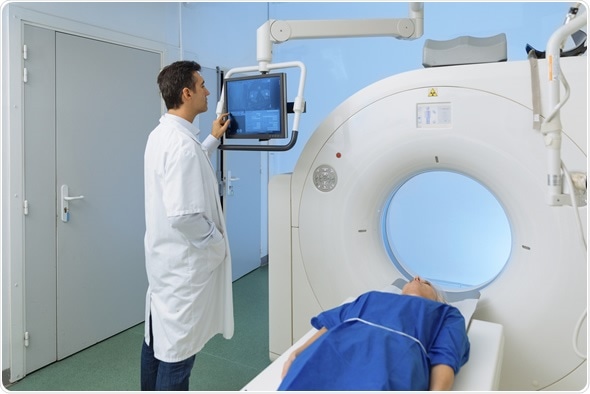
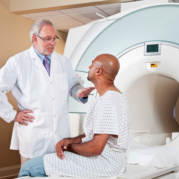
MRI technologists specialize in magnetic resonance imaging scanners. They inject patients with contrast media such as a dye so that the images will show up on the scanner. The patient is placed on a moveable bed that is inserted into the magnet.
Magnetic Resonance Imaging MRI is a non-invasive imaging technology that produces three dimensional detailed anatomical images. An MRI or magnetic resonance imaging is a radiology techinque scan that uses magnetism radio waves and a computer to produce images of body structures. The difference between an MRI and CT scan.
It is often used for disease detection diagnosis and treatment monitoring. Learn how to help your child prepare. An MRI scanner can be used to take images of any part of the body eg head joints abdomen legs etc in any imaging direction.
MRI is an imaging technique designed to visualise internal structures of the body using magnetic and electromagnetic fields which induce a resonance effect of hydrogen atoms. Unlike X-rays or computed tomography CT scans MRI does not use ionizing radiation. This exam does not use ionizing radiation and may require an injection of a contrast material called.
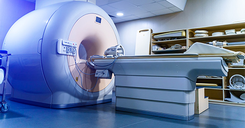
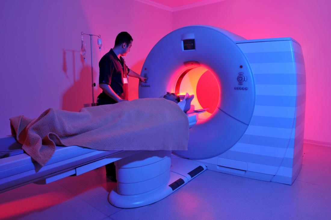
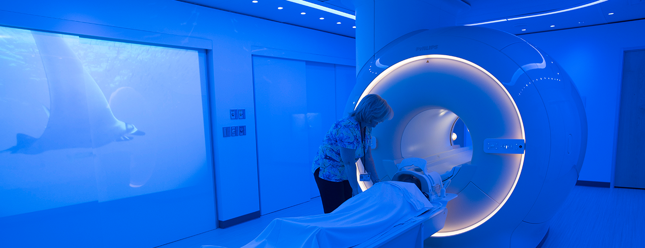
MAGNETIC RESONANCE IMAGING MRI Working as an MRI Technologist. The MRI scanner is a tube surrounded by a giant circular magnet. Magnetic resonance imaging MRI uses a powerful magnetic field radio waves and a computer to produce detailed pictures of the bodys internal structures that are clearer more detailed and more likely in some instances to identify and accurately characterize disease than other imaging methods.
Magnetic resonance imaging MRI is a medical imaging technique used to produce high quality images of the human body. Magnetic Resonance Imaging MRI Scans. In 2003 Paul C.
Unlike X-rays or CT images are created by using a magnetic field radio waves and a computer. In magnetic resonance imaging MRI k-space is the 2D or 3D Fourier transform of the image measured. Most MRI machines are large tube-shaped magnets.
Magnetic resonance imaging commonly called MRI is a method of looking inside the body without using surgery harmful dyes or X-raysInstead MRI scanners use magnetism and radio waves to produce clear pictures of the human anatomy. It was introduced in 1979 by Likes and in 1983 by Ljunggren and Twieg. The MRI machine is a large cylindrical tube-shaped machine that creates a strong magnetic field around the patient.
While supervised by board-certified radiologists MRI Technologists have responsibility and independence in performing their duties. MRI technology utilizes a powerful magnet to create a magnetic field that attracts and aligns hydrogen atoms inside the body. Lauterbur and Sir Peter Mansfield were awarded the Nobel Prize in Medicine for their discoveries concerning magnetic resonance imaging.
Some MRI machines look like narrow tunnels while others. No radiation is produced during an MRI exam unlike X-rays. MRI magnetic resonance imaging is a noninvasive diagnostic test that takes detailed images of the soft tissues of the body.
Magnetic Resonance Imaging MRI is an imaging technique designed to visualise internal structures of the body using magnetic and electromagnetic fields which induce a resonance effect of hydrogen atoms. An accurately timed sequence of radiofrequency. The magnet creates a strong magnetic field that.
The scanners use magnetic fields in combination with the contrast agent to produce images that a physician can use to diagnose medical problems. CT scans and MRIs are both used to capture images within your body. It allows your doctor to view your spine or brain in slices.
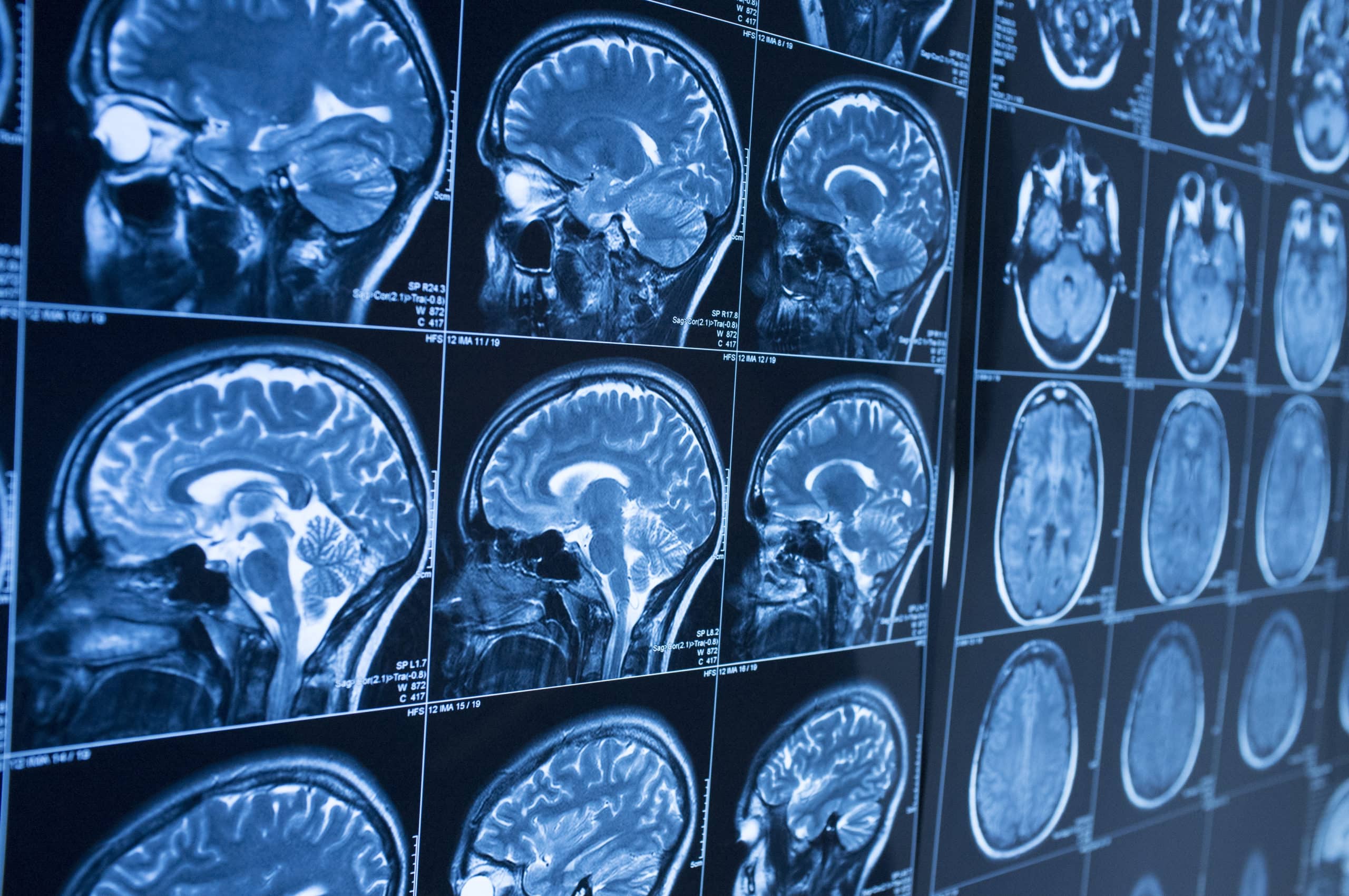
Learn more about this Canadian Medical Association accredited program. An MRI scan uses a large strong magnet combined with radio waves to generate multiple cross-section images that are then displayed on a computer. Magnetic resonance imaging MRI of the head uses a powerful magnetic field radio waves and a computer to produce detailed pictures of the brain and other cranial structures that are clearer and more detailed than other imaging methods.
MRI scanners create images of the body using a large magnet and radio waves. Journal of Magnetic Resonance Imaging JMRI is an international journal devoted to the timely publication of basic and clinical research educational and review articles and other information related to the diagnostic applications of magnetic resonance. In MRI physics complex values are sampled in k-space during an MR measurement in a premeditated scheme controlled by a pulse sequence ie.
The electromagnetic emission created by these atoms is registered and processed by a dedicated computer to produce the images of the body. The biggest difference is that MRIs magnetic resonance imaging use radio waves and. Magnetic Resonance Imaging MRI Technologists are valued members of todays healthcare team.
The physics of magnetic resonance imaging MRI concerns fundamental physical considerations of MRI techniques and technological aspects of MRI devices. Magnetic Resonance Imaging MRI is a unique tool that constructs cross-sectional pictures of internal organs and structures using radio waves and magnets. Magnetic Resonance Imaging MRI exams help physicians diagnose a range of conditions by producing images of internal organs and structures of the body.
It is based on sophisticated technology that excites and detects the change in the direction of the rotational axis of protons found in the water that makes up living tissues. MRIs provide a highly detailed picture of any part of the body and show a high level of detail of the. Magnetic resonance imaging MRI is a medical imaging technique that uses a magnetic field and computer-generated radio waves to create detailed images of the organs and tissues in your body.
Radio wave pulses are then focused on the aligned atoms in a. Estimating kinetic parameters from dynamic contrast-enhanced t 1-weighted MRI of a. MRI procedures play an important role in diagnosing diseases and injuries.
They used specialized MRI equipment to create images of structures inside the human body among other vital tasks. In our Magnetic Resonance Imaging MRI Graduate Certificate program learn how to you can start a career as an MRI technician or technologist in just eight months. Magnetic resonance imaging or MRI is a noninvasive medical imaging test that produces detailed images of almost every internal structure in the human body including the organs bones muscles and blood vessels.
It is used to evaluate the body for a variety of. How does an MRI work.
Magnetic Resonance Imaging Mri Overview

Magnetic Resonance Imaging Mri American College Of Veterinary Radiology

Magnetic Resonance Imaging Mri Ms Trust

Magnetic Resonance Imaging Mri
Basic Compartments Of The Magnetic Resonance Imaging Mri System Moore Download Scientific Diagram

Cardiac Magnetic Resonance Imaging Mri

Magnetic Resonance Imaging Mri Pleasantonimaging Com

Mri Cancerquest

Magnetic Resonance Imaging Mri Imaging Technology News

Magnetic Resonance Imaging Mri
Magnetic Resonance Imaging Mri Test Beaumont Health

Magnetic Resonance Imaging Mri Piedmont Virginia Community College
Mri Scans Definition Uses And Procedure
Magnetic Resonance Imaging Mri Mass Eye And Ear
Magnetic Resonance Imaging Mri Thanc Guide

How Long Does An Mri Magnetic Resonance Imaging Take

Magnetic Resonance Imaging Mri Private Hospitals Nsw
Magnetic Resonance Imaging Mri Exams Nuvance Health

Magnetic Resonance Imaging Mri Systems Market Set For Rapid Growth Of Usd 5 Billion By 2023 Asserts Mrfr With Ge Healthcare Philips Toshiba Corporation Xingaoyi Etc Medgadget
0 Response to "magnetic resonance imaging (mri)"
Post a Comment